SEC S20W1: | Hematopoiesis- Module 1
4 comments
 |
|---|
Hello friends and welcome to the SEC: S20/W1 in this great respect I would be carefully answering the questions to this respect and decisively taking part in the challenge. Here are my thoughts in this regard.
Anatomy and Functions of Hematopoietic Organs |
|---|
Hematopoiesis is actually the process of producing blood viable to be supplied to the various tissue organs and overall system in the body. It was coined from a Greek word Haîma which means blood and Poiēsis which means produce which definitely explain the technology.
From the lesson taught there are various Hematopoietic organs which are special in the body subject to time frame duration and effectiveness over a period of time.
Yolk sac: This is the first embryonic structure spotted during intra-uterine life in the foetus, discovered during the first week of embryonic life providing the embryo with intended nutrients, it is bag-shaped and indeed where hematopoiesis begins precisely at the 2nd week of embryonic life.
Liver: The liver is located at the right side of the abdomen, inferior to the thoracic cavity and superior to the abdominal cavity. This is the largest visceral organs found in man fashioned for overall metabolic activities in the body. It is a hematopoietic organ which begins shortly after the embryo develops into foetus in its intra-uterine life precisely, the first month of embryonic life until birth.
Spleen: This is a visceral organ found in the abdomen located superiorly at the anterior left abdomen which greatly function in engulfing and cell death of worn out blood cells and storage of blood. Its hematopoietic activity begins at the middle of second month of embryonic life up until the seventh month.
Thymus: This is a smaller organ located anteriorly at the superior part of the thoracic cavity lying anteriorly to the lymph nodes, responsible for the proliferation of blood cells, it's life span runs from after foetal life, birth up until puberty
Bone marrow: This is a sponge-like tissue which is located at the anterior and posterior end of most bones prevalently at the forelimb and hindlimb or joints, together with the synovial fluid. It produces blood cells in man and gradually becomes the major producer of blood cells in adult human.
Medullary Hematopoiesis vs Extramedullary Hematopoiesis |
|---|
Medullary Hematopoiesis is basically production of blood cells in the bone marrows precisely. These bone marrows are prevalent in the long bones of the body, basically the forelimb, hindlimb, pelvic girdle, just to mention but a few.
Extramedullary Hematopoiesis occurs when there is production of blood cells asides the bone marrow, post embryonic life. This occurs in the spleen, liver and mostly as a support during condition where the body undergoes physiological backlogs due to infections, age amidst other factors.
Diagram Of Hematopoiesis with explanation |
|---|
 |
|---|
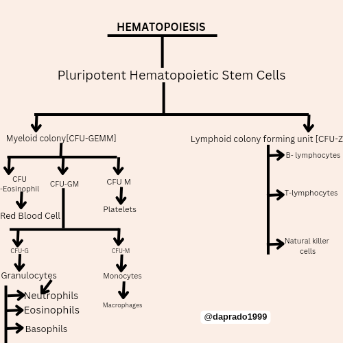
The Pluripotent Hematopoietic Stem Cells are divided into the myeloid colony forming unit [CFU-GEMM] and lymphoid colony forming unit [CFU-L] The myeloid colony further divides into the [CFU-E] [CFU-GM] and [CFU-M] (which later divides into the platelets). The Lymphoid colony forming unit [CFU-L] further divides into B-lumphocytes T-lymphocytes and Natural Killers which are body fighters of the white blood cells. The [CFU-E] further forms the red blood cells. The [CFU-GM] further divides forming the [CFU-GM] and [CFU-M] The [CFU-G] further divides forming Granulocytes which further divides into the Neutrophils Eosinophils and Basophils The [CFU-M] forms the Monocytes and Macrophages this certainly explains the Hematopoietic process.
Clinical Care |
|---|
A 25-year-old male patient comes into the emergency room of the Ruiz y Páez University Hospital Complex with bruises all over his body from a car crash. He is given an order for tests requesting a complete blood count, blood glucose, urea, creatinine, glomerular filtration rate, and lipid profile. The test results show significant blood loss and kidney problems. X-rays are also performed showing a broken femur. Explain the following in your own words:
Blood flow as a result of ruptured tissues which caused blood flow profusely and subsequently became the reason for blood loss. The blood loss also shows that the platelets clotting activities in the blood cell was shot which means it was a major rupture which could damage the victim.
The fracture to the femur poses threat because it is the basic point of blood formation, this is the overflow of blood into the blood stream during to internal bleeding could be a reason for blood loss.
Sincerely, the donor would be a young universal donor with the O blood group which is neutral and does not affect the antibodies and antigens of the blood group. Highly essential and compatible. It would be best to donate to the victim from the universal donor.
Comments